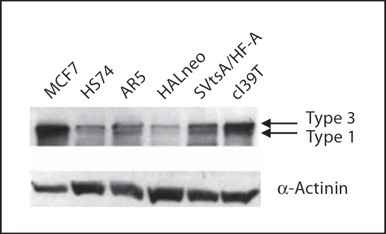
Fig. 7.

Expression of MLLT4 in SV40-immortalized human cells. 30 μg of the protein of each cell line was loaded and electrophoresed in 7.5% denaturing SDS polyacrylamide gel. After protein transfer the blot was probed with MLLT4-specific antibody and with the secondary antibody to visualize MLLT4 proteins. The same blot was probed with anti-α-actinin antibody for loading control. See text for different cell line designations. It is difficult to distinguish the larger transcripts of MLLT4 from each other. MCF7, a breast epithelial cancer cell line, was used as a positive control.
