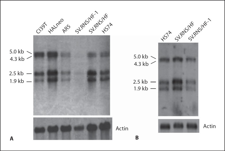
Fig. 8.
Expression analysis of PHF10 in normal, preimmortal and SV40-immortalized cell lines. A 3 μg of polyA+ RNA from the indicated cell lines was fractionated in 0.8% sodium borate-formaldehyde agarose gels. After blotting, the membrane was hybridized with 32P-labeled 500-bp fragment of PHF10b cDNA from the 5′ end using PerfectHyb Plus hybridization buffer (Sigma). After washing, the membrane was exposed to X-ray film for 3 h. B Northern blot of the indicated cell lines probed as in panel A except that polyA+ RNA was fractionated in 1% sodium borate-formaldehyde agarose gels. The polyA+ RNAs were prepared independently in panel A and B.

