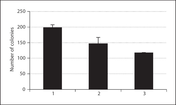
Fig. 9.
Effect of overexpression of PHF10b(HA) in cl39T/Tet-On cells as monitored by colony formation assay. 600 cells of different cell lines were plated on 100-mm dishes in 3 replicates. Cells from a stable clone carrying PHF10b(HA) under the control of pTRE-tight promoter were either treated with DOX (1 μg/ml) to induce PHF10b(HA) expression or kept in culture medium containing Tet-free serum as a control. Cells were fed every 3 days with medium containing DOX or without DOX. After 21 days, colonies were fixed, stained with crystal violet and counted. Data presented are the mean of 3 independent experiments. Column 1 – cl39T Tet-on cells, column 2 – cl39T/Tet-On·PHF10b(HA) cells (DOX–), and column 3 – cl39T/Tet-On·PHF10b(HA) cells (DOX+).

