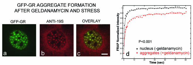
FIG. 6.
Nuclear GFP-GR aggregates. (a to c) Geldanamycin treatment combined with stress (heat, cold, or prolonged imaging) causes the disappearance of arrays and the formation of GFP-GR spots, which colocalize with a proteasome antibody. (d) In these spots, a fraction of GFP-GR is immobilized compared to other regions of the nucleus. During imaging, these aggregates appear more rapidly with corticosterone than with dexamethasone. Scale bar, 5 μm.

