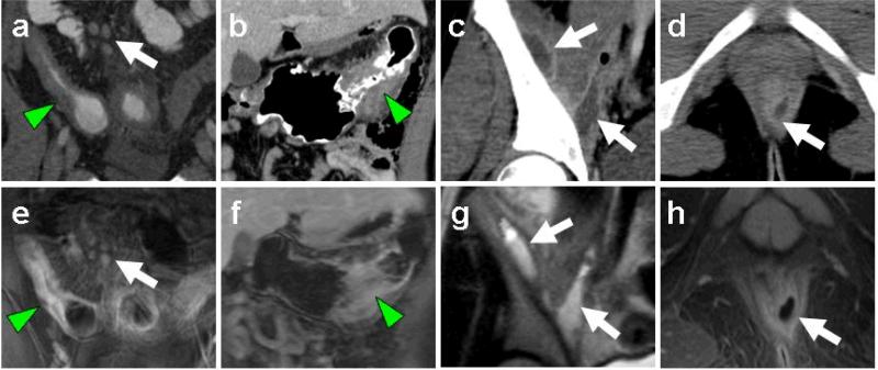
Fig. 1.
MR enterography and CT depiction of Crohn's imaging features. Representative image pairs from CT (a-d) and MR-E (e-h) studies demonstrate wall thickening of small bowel (a, e arrowheads) and colon (b, f arrowheads), lymphadenopathy (a, d arrows), intersphincteric fistula formation (d, h arrows), and iliopsoas abscesses (c, g arrows).

