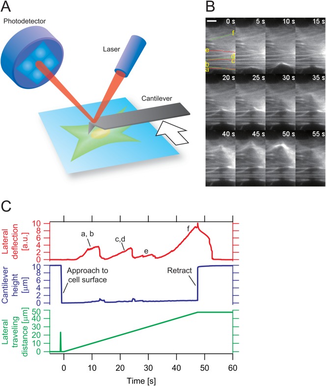
Fig. 1. Creation of localized mechanical damage on SFs by AFM manipulation.
(A) Experimental design for the application of lateral force to SFs. The rat fibroblast cell line, VNOf, transiently coexpressing AcGFP1-actin and TagRFP-focal adhesion protein, was cultured on fibronectin-coated glass slips. The probe tip of the AFM cantilever pushed the cell in close proximity to one of the fluorescently visualized SFs. The cantilever was then moved sideways, pushing and straining the SFs in the lateral direction. (B) Time-lapse micrographs of SFs before (0 s) and during (5–55 s) lateral force application. The major SFs (a–f) were strained by lateral travel of the probe tip. The entire process was also recorded as lateral deflection of the cantilever (C). The strained SFs underwent thinning from the damaged sites. Scale bar: 10 µm.

