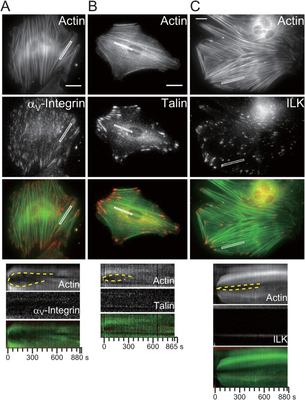
Fig. 3. The protein complex which repairs damaged SFs is not a novel focal adhesion.
The micrographs and kymographs are of AcGFP1-actin and TagRFP-focal adhesion proteins in rat fibroblasts (A: αV-integrin, B: talin, C: ILK). The kymographs (bottom) were obtained from the white boxes in the micrographs. The dashed yellow lines in the kymographs show the strained SFs undergoing thinning, elongation, and then repair. Scale bars: 20 µm.

