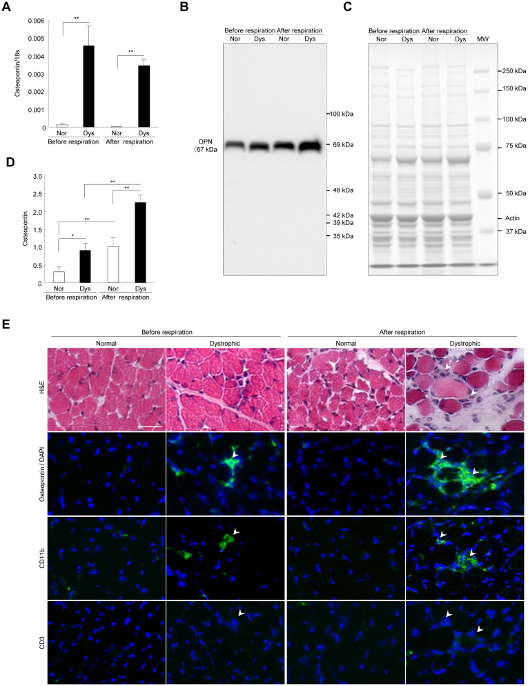
Figure 3. Osteopontin upregulated before the diaphragm damage in neonatal dystrophic dogs.
(A) Comparison of relative osteopontin mRNA levels to 18 s in normal (Nor) and dystrophic (Dys) dogs before (n = 4, each) and after (n = 4, each) the respiration. Bar: mean ± SD; ** p < 0.01. (B) Western blotting of osteopontin and (C) CBB staining of diaphragms in normal and dystrophic dogs before and after respiration. Αctin: loading control. The short and long exposure blots are included in the supplementary information. (D) Comparison of relative levels of 69 kDa osteopontin to actin in normal (Nor) and dystrophic (Dys) dogs before (n = 4, each) and after (n = 4, each) the respiration. Bar: mean ± SD; ** p < 0.01. (E) Hematoxylin-eosin (H&E) staining of diaphragms of normal and dystrophic dogs before respiration and 1 hour after respiration. Immunohistochemistry of osteopontin, CD11b, CD3 (all green), and DAPI (blue) in normal and dystrophic dogs before and after respiration. Bar indicates 100 μm.

