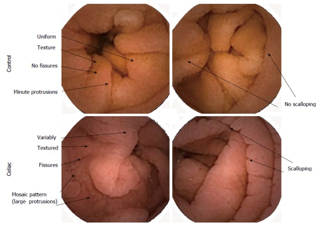
Figure 1.

Color images from videocapsule device. Differences in celiac images where villous atrophy is present are shown vs control. Control images have a more uniform texture at the mucosal surfaces. Celiac surfaces have a rougher appearance, with more fissuring, large protrusions, and scalloping along the folds.
