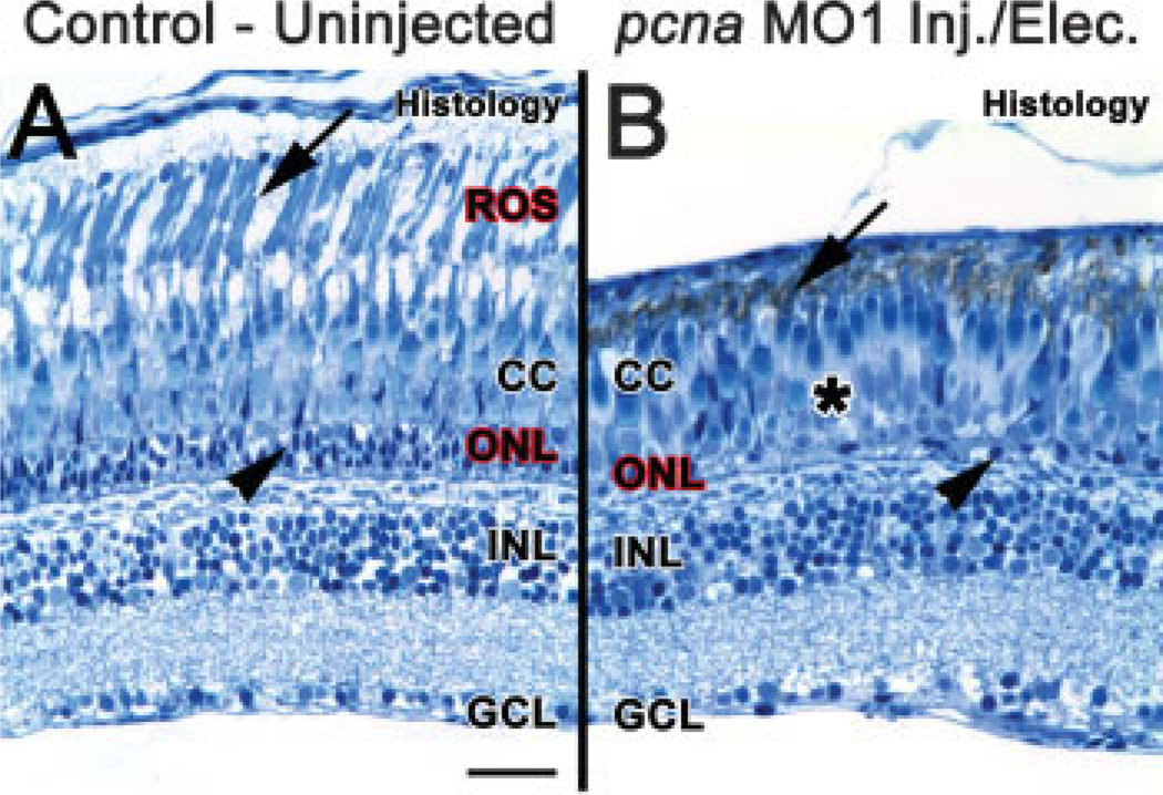
Figure 7.
PCNA knockdown results in a reduction of ROS and ONL nuclei 28 days post light treatment. At 28 days following constant light treatment, histological sections of uninjected retinas (panel A) showed a regenerated outer nuclear layer (ONL, arrowhead), cone cell layer (CC) and rod outer segments (ROS, arrow). In contrast, histological sections from pcna morphant retinas (panel B) showed an absence of distinguishable ROS (arrow), very few nuclei present in the ONL (arrowhead), a disorganized cone cell layer, and large sections absent of long single cones (asterisk). Scale bar: panel A, 25 µm (A–B).

