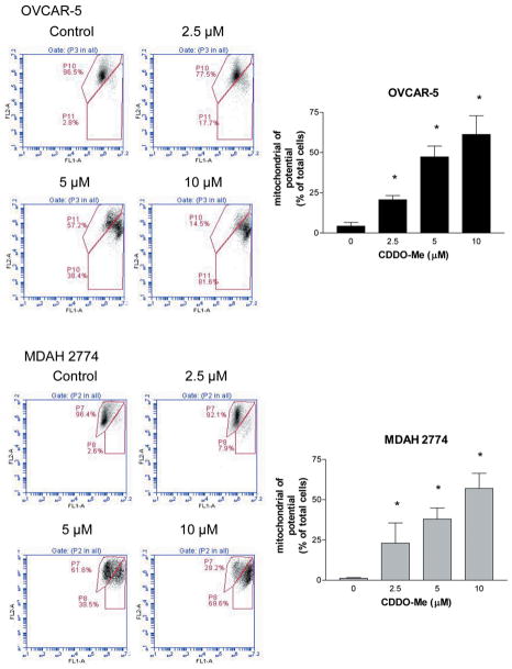
Figure 4.
CDDO-Me induces mitochondrial depolarization. OVCAR-5 and MDAH-2774 cells were treated with CDDO-Me at 0 to 10 μM for 20 h. Cells were loaded with mitochondrial potential sensor JC-1 (10 μg/ml) for 10 minutes at 22°C and analyzed by flow cytometry for cells fluorescing red (FL2 channel) or green (FL1 channel) (A). Bar graphs show the percentage of cells with loss of mitochondrial potential (B). Similar results were obtained in two separate experiments. *p<0.05 compared to control cells (no CDDO-Me).

