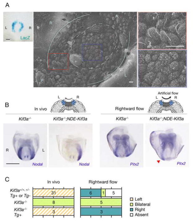
Figure 5. Nodal flow is sensed by the cilia of crown cells.
(A) An E8.0 transgenic embryo harboring NDE-Kif3a-IRES-LacZ was stained with X-gal, revealing crown cell specific expression of the transgene (left). Scale bar, 50μm. In scanning electron microscopy of the node of an E8.0 Kif3a−/− embryo with NDE-Kif3a, cilia are apparent at the edge (boxed by the red line), but not at the center (boxed by the blue line) of the node. Pale blue lines indicate the border between the endoderm and crown cells, with the dotted circle enclosing pit cells. The boxed regions on the left are shown at a higher magnification on the right. Scale bars, 5 μm. (B) Expression of L-R marker genes in embryos of the indicated genotypes that were examined at E8.0 (in vivo) or cultured under the influence of a rightward artificial flow before analysis. Note that Pitx2 expression pattern of the Kif3a−/−;NDE-Kif3a embryo responded to the flow and is right-sided (red arrowhead). Scale bar, 500 μm. (C) The numbers of embryos showing each pattern of gene expression are summarized.

