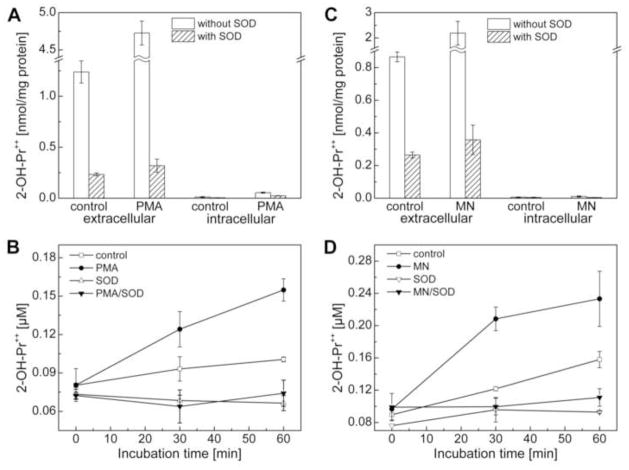
Figure 10.
Measurement of extracellular superoxide radical anion production using the HPr+ probe. (A) RAW macrophages were stimulated with PMA (1 μM) in cell culture media containing HPr+ (60 μM) (as described in the Materials and Methods section) in the presence and absence of SOD (0.1 mg/ml) for 1 h min and 2-OH-Pr++ levels were measured in cell culture media (extracellular) and cell lysates (intracellular). To directly compare the amount of 2-OH-Pr++ inside the cell or media, the amount of the product was normalized to the total amount of protein in the cell lysates. (B) Time course measurements of extracellular 2-OH-Pr++ under different experimental conditions, as indicated. Experimental conditions: PMA (1 μM), HPr+ (60 μM) and SOD (0.1 mg/ml). (C) Same as (A) except that superoxide production was stimulated by adding menadione (100 μM) and incubated for 30 min. (D) Same as (B) except that macrophages were treated with menadione (MN, 100 μM) instead of PMA.

