
FIG. 7.
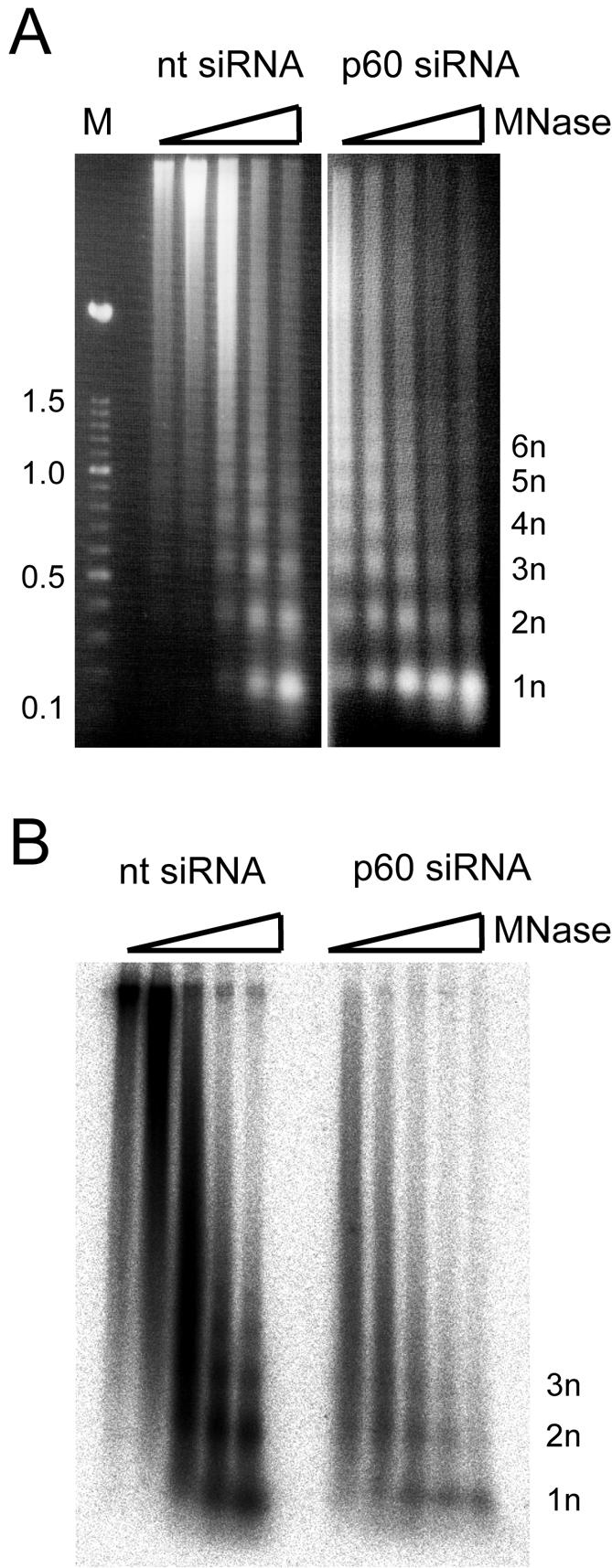
Analysis of chromatin structure in proliferating cells during silencing of p60. HeLa cells were transfected with either nontarget (nt) or p60 siRNAs, and a cell homogenate of each sample was prepared at 24 h posttransfection. Nascent DNA in these nuclei was radiolabeled for 3 h in vitro (see Materials and Methods). Labeled nuclei were treated with 0.1, 0.3, 1, 3, and 10 U of micrococcal nuclease (MNase) per ml (lanes 1 to 5, respectively). Nuclease-resistant DNA from both transfection experiments was isolated and analyzed by agarose gel electrophoresis. Note that DNA prepared from nontarget and p60 siRNA-transfected cell nuclei did not enter the gel after incubation without micrococcal nuclease present and is therefore not shown here. (A) Visualization of digested bulk DNA from transfected cells by staining with ethidium bromide. Lane M, multimeric 100-bp DNA ladder (Roche), with size indicated in kilobase pairs. The positions of nuclease-resistant mono- (1n) and oligonucleosomal DNA fragments are indicated. (B) Visualization of digested replicating, nascent DNA in these nuclei by phosphor imaging of the dried gel.