
Figure 1.
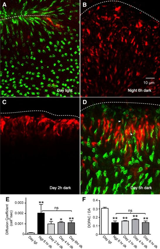
Photoreceptor coupling and dopamine release in light- and dark-adapted mouse retina in daytime and at night. Cut loading was conducted in mouse retina using Neurobiotin (red; A–D) followed by postfixation labeling of cones with PNA (green; A, D). Images show flat-mounted retina at the plane of the photoreceptor inner segments. Under light exposure in the daytime (A), tracer diffusion was limited to the cut edge (dashed line). The majority of Neurobiotin-loaded cells were not colocalized with PNA. In the nighttime 6 h dark-adapted retina (B), Neurobiotin tracer diffusion was much more extensive among the photoreceptor matrix. Daytime photoreceptor coupling was increased by dark adaptation for 2 h (C), 4 h, and 6 h (D), with a small fraction of cones among the coupled cells (green PNA label in D). E, Summary of tracer coupling data for day; night; and 2, 4, and 6 h dark adaptation in the daytime (n = 5–11 per condition). F, HPLC analysis of dopamine and DOPAC in whole eyes from animals collected under the same conditions as for tracer coupling measurements (n = 4–6 per condition). Data in E and F are mean ± SEM; *p < 0.05; **p < 0.01; ns not significant. A and D are projections comprising 4 μm focal depth; B and C are projections comprising 0.9 μm focal depth.