
Figure 7.
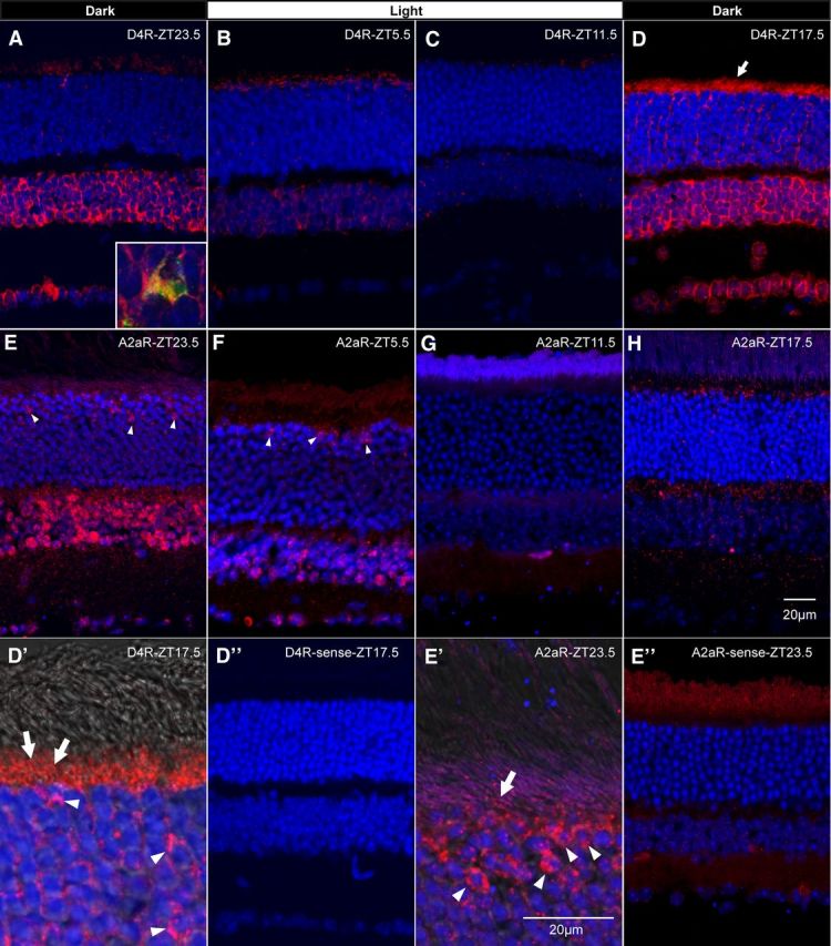
ISH for dopamine D4R and adenosine A2aR in mouse retina at different time points during an L/D cycle. A–D, D4R mRNA signal (red) was located in photoreceptors, most INL neurons, and ganglion cells. The sense probe resulted in negative labeling at all time points (ZT17.5 shown in D″). Some cells in the INL showed positive tyrosine hydroxylase antibody colabeling (green; inset in A). DAPI was used as nuclear counterstain (blue). The D4R transcript level was high at night (A, D) and low during the day (B, C). E–H, A similar distribution pattern was observed for the A2aR transcript (red), with signal observed in the upper ONL (arrowheads), most cells in the INL, and some ganglion cells. Sense probe labeled negatively at all time points (ZT23.5 shown in E″). The A2aR transcript seemed high before light onset and during mid-day. Interestingly the A2aR mRNA was mainly located surrounding the somata in the upper ONL (arrowheads in E, E′, and F), with some mRNA signal in the inner segments (arrow in E′). In contrast, the majority of D4R mRNA was located in the inner segments (arrows in D and D′), with minor signal surrounding photoreceptor somata (arrowheads in D′). Merged images combining bright field and fluorescence microscopy to provide image of photoreceptor outer segments (D′, E′).