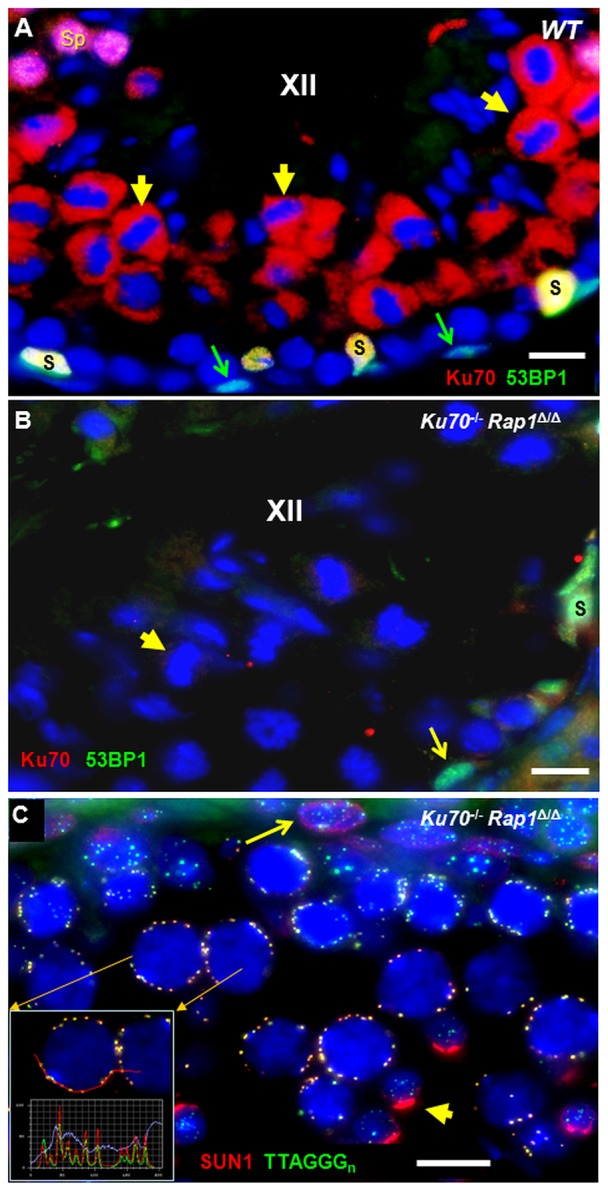
Fig. 2.

Ku70 (red) and 53BP1 (green) immunofluorescence (IF) staining pattern of wild-type (WT) and Rap1Δ/ΔKu70−/− testes tubules. (A) WT stage XII tubule showing Ku70 protein IF (red) in the cytoplasm of metaphase I and II cells (examples highlighted by short yellow arrows) near the tubule center. Sertoli cells (S) at the tubule periphery appear yellow owing to colocalization of red Ku70 and green 53BP1 fluorescence. A-type spermatogonia show strong greenish 53BP1 expression (green arrows). Spermatids (Sp) appear pink owing to strong fluorescence for Ku70 and 53BP1 at the upper left corner of the image. (B) Ku70−/− stage XII tubule showing absence of red Ku70 protein signals (example indicated by short yellow arrow), whereas an A-type spermatogonium (long arrow) and a Sertoli cell (S) show green 53BP1 fluorescence. (C) IF of SUN1 (red) and (TTAGGG)n telomere FISH (green) revealing the colocalization of SUN1 with meiotic telomeres at the nuclear periphery of pachytene spermatocytes in Rap1Δ/Δ Ku70−/− testis tissue section. The inset shows fluorescence profiles across several telomeres that highlight colocalizing SUN1 (red line) and telomere (green line) fluorescence peaks. This staining is identical to that in wild type (not shown) (cf. Scherthan et al., 2011). A punctate NUP-like somatic distribution pattern of SUN1 (red) is seen at the meiotic NE of a spermatogonium at the tubule periphery (arrow), whereas haploid spermatids (short yellow arrow) display a strong red acrosomal signal. Scale bars: 10 µm.
