Fig. 3.
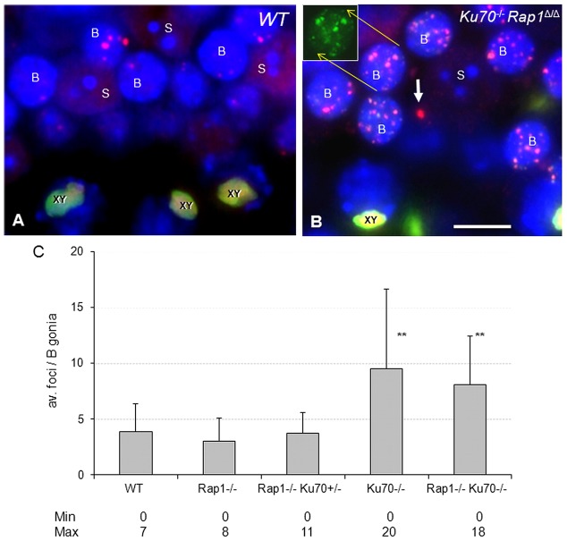
DNA damage in B spermatogonia of Ku70-deficient testes revealed by 53BP1 (red) and γH2AX staining (green). (A) Testicular section of a wild-type mouse showing a few 53BP1 foci (red) in B spermatogonia (indicated by letter B). Wild-type Sertoli cells (S) show no 53BP1 foci. XY denotes the 53BP1- and γH2AX-positive sex body (yellow) of pachytene spermatocytes. (B) Numerous 53BP1 DNA damage foci in B spermatogonia of a Ku70−/−Rap1Δ/Δ testis section. These colocalize with γH2AX foci (green) as shown by the green channel image of one nucleus in the inset. The white arrow denotes a large 53BP1 DNA damage focus in the DAPI-faint chromatin of a double knockout Sertoli cell nucleus. (C) γH2AX and 53BP1 foci numbers are significantly increased (**P<0.001) in Ku70-deficient B spermatogonia of single and double knockout testes relative to the Rap1Δ/Δ, heterozygous and wild-type testes. Error bars indicate s.d.

