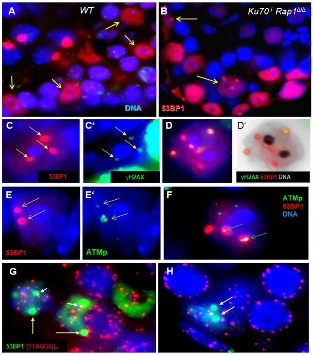
Fig. 7.
Persistent DNA damage and active DDR in Ku70-deficient Sertoli cells of testes sections. (A) Wild-type Sertoli cells (arrows) show strong diffuse 53BP1 fluorescence (red) throughout the nucleus except for the nucleolus (dark dot) at its center. Bright red dots in spermatocyte nuclei represent the XY body. (B) Ku70−/−Rap1Δ/Δ Sertoli cells displaying large red 53BP1 DNA damage foci (arrows) in their nuclear chromatin. The two blue dots next to the dark spot (the nucleolus) represent the chromocenters that are specific for this cell type. (C-D′) Ku70-deficient Sertoli cells showing colocalization of strong 53BP1 (red) with weaker γH2AX signals (green) at large DNA damage foci (arrows). (D) Sertoli cell with six 53BP1 foci, four of which contain γ-H2AX fluorescence (green) as indicated by their yellowish color. (D′) Same cell as in D with the blue color of the DAPI stain converted to gray for better display. (E,E′) Colocalization of 53BP1 (red) with activated ATM (ATMp, green) at two large DNA damage foci (arrows) of a Ku70−/− Sertoli cell. One focus (lower arrow) contains a large amount of ATMp. (F) Sertoli cell with three damage foci, two of which contain ATMp (arrows). (G,H) TTAGGGn telomere FISH signals (red) only rarely colocalize with 53BP1 damage foci (green; arrows) in Ku70−/− Sertoli cell nuclei. In such cases, the small telomere signals lie at the border of the large 53BP1 foci (short arrows).

