Fig. 8.
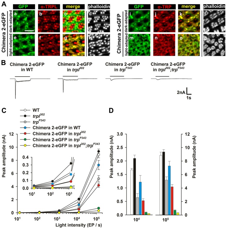
Chimera-2–eGFP predominantly interacts with the TRP channel. (A) Subcellular localization of chimera-2–eGFP, TRPL and TRP in dark-adapted (16 hour) and light-adapted (16 hour orange light) Drosophila eyes, expressing chimera-2–eGFP in the WT background. Fluorescence microscopy of eye cross sections, showing fluorescence of eGFP-tagged channels (green), immunofluorescence of anti-TRPL (left panel, red) or anti-TRP antibodies (right panel, red) and phalloidin labeling of the rhabdomeres (white). A merge of the green and red fluorescence is also shown. Scale bar: 10 µm. (B) Representative responses of chimera 2 in the WT, trpl302, trpP343 and trpl302;trpP343 backgrounds to 1.3×10⁵ EP/s. (C) Intensity–response (R-logI) relationship of the fly strains as in A (n = 5, means ± s.e.m.). Inset: intensity–response relationship in dim light (n = 5, means ± s.e.m.). (D) Histogram plotting the peak amplitude of the response to 1.3×10⁴ EP/s (left) and 1.3×10⁵ EP/s (right) of the fly strains as in B.

