
Fig. 3.
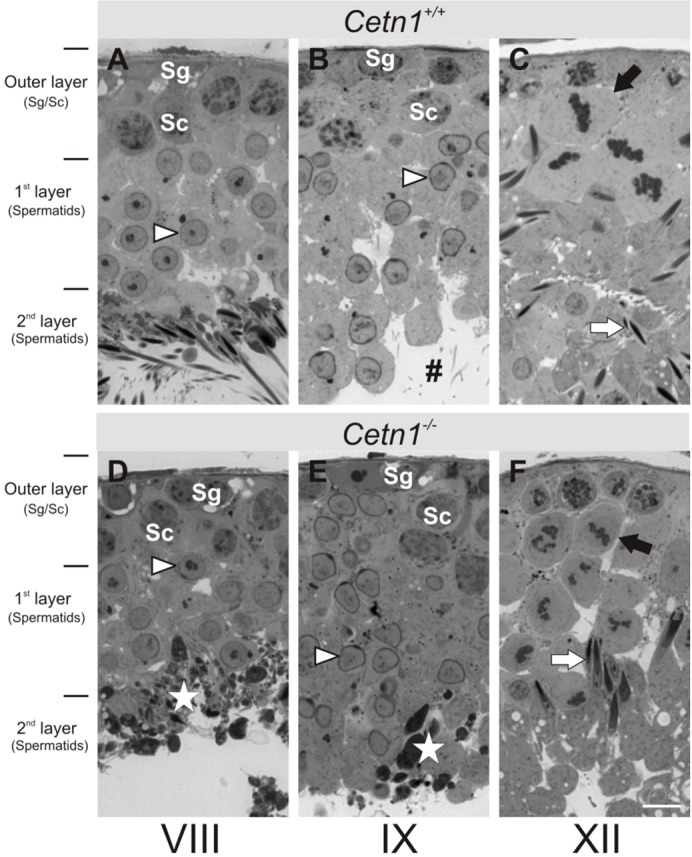
Seminifereous tubules of Cetn1+/+ and Cetn1−/− mice. Stage-matched seminifereous tubules from (A–C) Cetn1+/+ mice and (D–F) Cetn1−/− mice. The outer layer consists of spermatogonia (Sg) and spermatocytes (Sc), the first layer consists of spermatids (arrowheads) from round Golgi phase to the end of acrosome phase; the second layer harbors the maturating spermatids up to spermiation into the lumen of seminifereous tubules. (A) Stage VIII tubules of Cetn1+/+ mice with spermatocytes in the outer layer, round spermatids in the first layer and tailed spermatids in the second layer at the luminal side of the tubules. (B) Stage IX tubules of Cetn1+/+ mice. Instead of round spermatids, capped spermatids entering acrosome phase are present in the first tubule layer. #, Tails of mature spermatids after spermiation in the tubule lumen. (C) Stage XII tubules of Cetn1+/+ mice exhibit spermatocytes at meiosis (black arrow) in the first layer forming the new generation of spermatids. Elongated spermatids (white arrow) are present in the second layer. (D) Stage VIII tubules of Cetn1−/− mice with spermatocytes in the outer layer, and round spermatids in the first layer. Tailed spermatids in the second layer are absent, but dark, dense material of degraded spermatids (white star) is present. (E) Stage IX tubules of Cetn1−/− mice reveal the same layering as Cetn1+/+ mice, but contain dark dense material of degraded spermatids (white star). (F) Stage XII tubules of Cetn1−/− mice show spermatocytes during meiosis (black arrow) in the first layer and elongated spermatids at the transition to maturation phase (white arrow). Layering of the seminifereous tubules was the same in Cetn1+/+ and Cetn1−/− mice. Scale bar: 10 µm.