
Fig. 5.
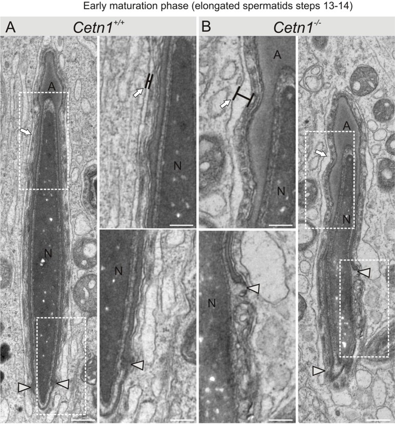
Ultrastructure of early maturation phase spermiogenesis of Cetn1+/+ and Cetn1−/− spermatids. (A,B) The first differences between Cetn1+/+ and Cetn1−/− were found in steps 13–14. The caudal movement of the acrosome forming the acrosomic clefts (arrowheads) was uneven and a general bulky appearance was observed in Cetn1−/− spermatids. Sertoli cell membranes were deformed compared to the straight, parallel membranes of Cetn1+/+ mice (arrows). At higher magnification (center two panels), wider distention between the Sertoli cell membranes was seen (arrows, black bars). Also at high magnification, at the spermatid base a disruption to the caudal migration of the acrosome was evident (B, arrowheads). Scale bars: 0.5 µm.