
Fig. 6.
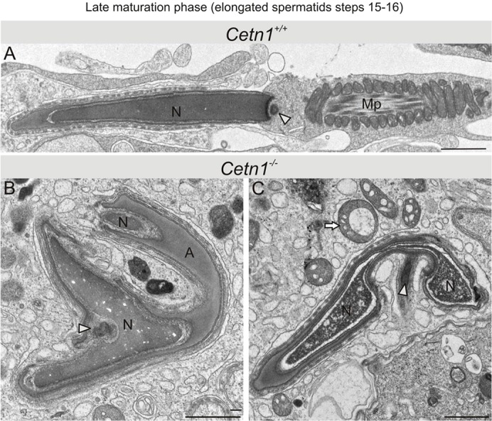
Late maturation phase spermatids. Spermatids were analyzed by high resolution electron microscopy at steps 15–16 of spermiogenesis. (A) Wild-type spermatid showing a midpiece (Mp), an elongated nucleus (N) and a remaining centriolar structure (arrowhead). (B,C) Abnormalities were prominent in Cetn1−/− spermatids. No midpiece or tail formation was found. Nuclei were split, bifurcated and the chromatin appeared perforated. Additionally, spermatid heads were bent over and the shape of the centrosomal region (arrowheads) was abnormal. Cell debris of spermatids in granules (C, arrow) was observed, indicating cell degeneration. A, acrosome. Scale bars: 1 µm.