
Fig. 8.
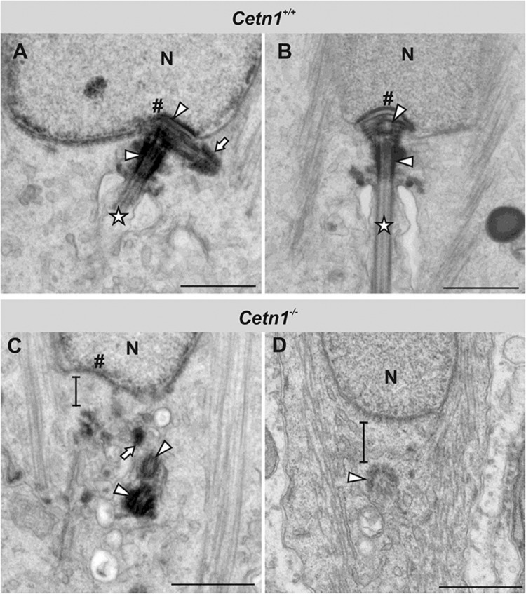
Ultrastructure of spermatid centriole rearrangement during late spermiogenesis. (A,B) Cetn1+/+ spermatids during centriole rearrangement of late spermiogenesis. (A) Proximal and distal centrioles (arrowheads) positioned directly adjacent to the fossa (#) of the nucleus (N). The proximal centriole projects the adjunct (arrow), and the sperm flagellum axoneme is visible (star). (B) Rearranged centrioles (arrowheads) finish to form the basal body apparatus at the nuclear fossa (#), from which the sperm flagellar axoneme extends (star). (C,D) Cetn1−/− spermatids during centriole rearrangement. (C) Both centrioles (arrowheads) and the adjunct remnant (arrow) appear disengaged from the nuclear membrane, as indicated by the black bar. The nuclear fossa (#) is dysmorphic. (D) The distal centriole (arrowhead) projects a tubular structure corresponding to the axoneme, but it is bent and flaccid. Instead of a modified proximal centriole, a cytoplasmic gap (indicated by black bar) appears between the nucleus (N) and the distal centriole. Scale bars: 0.5 µm.