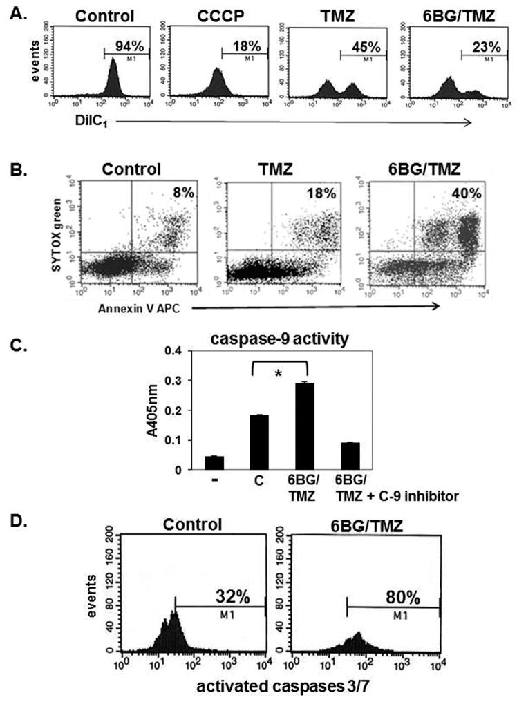
Figure 3. Analysis of mitochondrial membrane depolarization and apoptosis following exposure to myelosuppressive chemotherapy.
Human MP cells were treated with 6BG +/- TMZ for 4 days. (A) Mitochondrial membrane depolarization was determined by DiIC1 staining. Increased depolarization correlates with decreased DiC1 stain. Insert on control histogram is DiIC1 staining of myeloid cells treated with the membrane depolarization agent, CCCP. (B) Apoptotic cell death was determined by Annexin V and Sytox- green stain. (C) Caspase-9 activity was determined in the +/- of the caspase-9 specific inhibitor. (D) Caspase-3/7 activation was determined by flow cytometry. Data are representative of 3 independent experiments for A-C and 2 independent experiments for D.

