
Figure 1. Tumor growth and enlarged spleens in immune competent BALB/c mice.
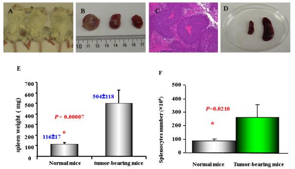
Six to eight weeks old BALB/c mice were injected (s.c.) 1MEA cells ( 5 × 10⁶ / per mouse). After two weeks, the tumors were measurable. As mentioned in Materials and Methods, at the endpoint of experiments (the end of the forth week), spleens and tumor tissues were collected and weighed. Splenocytes were prepared and counted after red cells lysis. A. Tumor-bearing BALB/c mice; B. Tumor tissue separated from the mice. C. Histological features of the liver cancer tissue (H&E stain) showing poorly differentiated hepatocellular carcinoma; D. the size of spleens from Balb/c mice with tumor (right) and normal mice (left); E. the weight of spleens from normal mice and tumor-bearing mice (n=9); F. the splenocytes from normal mice (87± 14 × 106; n=3) and tumor-bearing mice (263 ± 95 × 106; n=5). Data represent the mean ± SD from one of two similar experiments