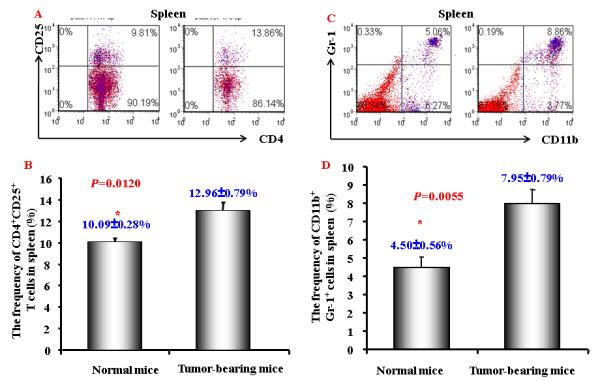
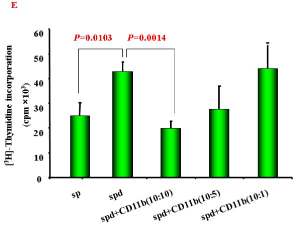
Figure 2. Increased CD4+CD25+ T cells and CD11b+Gr-1+ cells in tumor-bearing mice.


A. A representative flow cytometric data showing the CD4+CD25+ T cells frequency of gated CD4+ T cells in splenocytes from normal and HCC tumor-bearing mice; B. Mean CD4+CD25+ T cells frequency in CD4+ T cells in splenocytes from normal and HCC tumor-bearing mice (n=3) . C and D. A representative (C) and mean ( D) flow cytometry data showing increased CD11b+ Gr-1+cells frequency in splenocytes from normal and HCC tumor-bearing mice (n=3). The gated viable cells were analyzed. Numbers in the figures represent the percentage of fluorescence-positive cells in corresponding areas; E. Functional analysis of CD11b+ cells were evaluated for their ability to suppress autologous CD11b+-depleted splenocytes proliferation by [3H]-thymidine incorporation when stimulated with anti-CD3 and CD28 at various ratios. Values represent mean ± SD (n=3) and are representative examples of three separate experiments.
