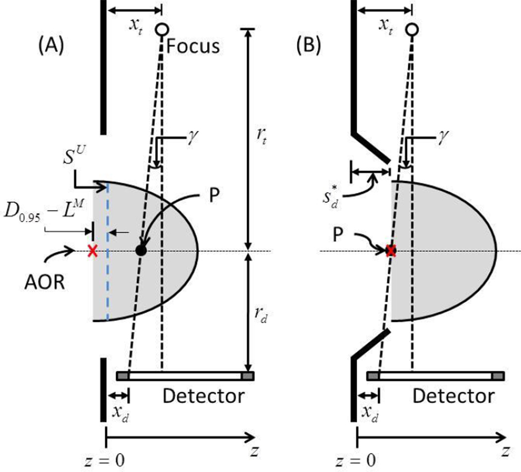
Figure 9.
Illustration of the method used to determine the optimal swale-depth in upright breast CT (not drawn to scale). The barrier used in the study had a flat surface. SU is the skin mark with the subject positioned for upright breast CT and is aligned with the anterior surface of the barrier. D0.95 − LM is the amount of breast tissue posterior to SU that needs to be imaged so as to obtain equivalent chest-wall coverage in breast CT images as mammograms for 95% of the subjects and is marked by a cross. γ is the cone angle subtended by a ray from the x-ray focal spot to the first row/column of the detector and intersects the axis of rotation at the point marked P . In (B), the swale depth needed so that the breast is shifted in the anterior direction such that the point marked by the cross is congruent with P is shown.

