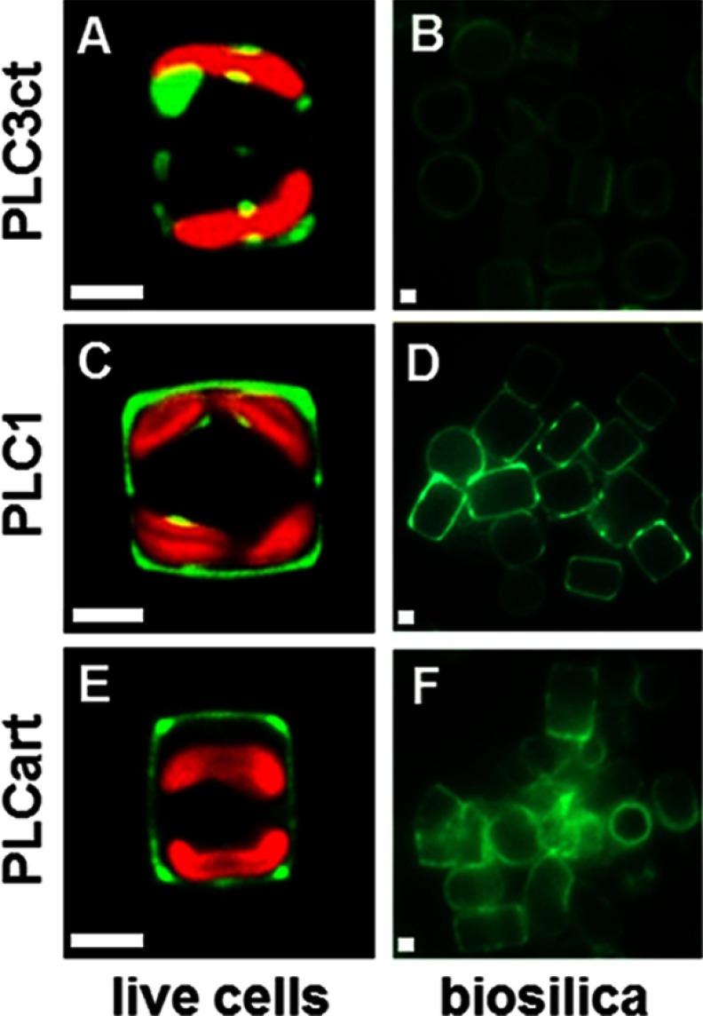
FIGURE 6.
Silica-targeting efficacy of pentalysine clusters. A, C, and E, confocal fluorescence microscopy images of individual live cells in girdle view expressing the indicated pentalysine cluster-GFP fusion proteins. B, D, and F, epifluorescence microscopy images of isolated biosilica from multiple cells of the same transformant strains. Green color is indicative of Sil3-GFP, and red color is caused by chloroplast autofluorescence. White bars: 2 μm.

