
TABLE 2.
MicroCT analysis showed increased trabecular and cortical bone in 12-week-old Ocy-PPRKO females
Trabecular bone parameters were in L5 vertebrae. Cortical bone parameters were in the midshaft of femurs. Values are mean ± S.E., two-tailed t test assuming equal variance.
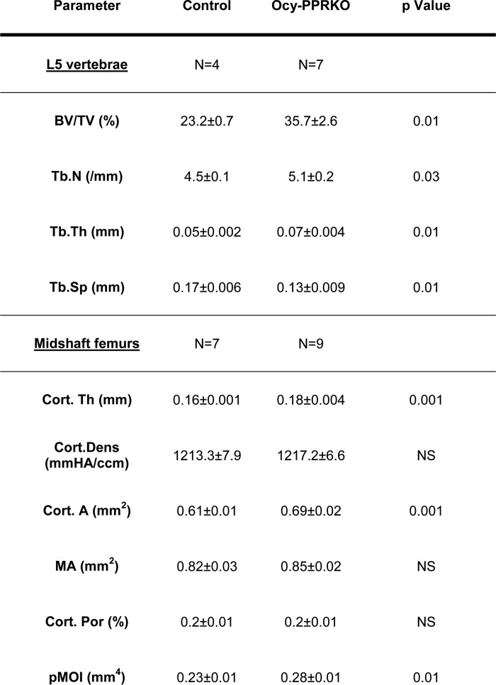
* p ≤ 0.05, and NS = not significant.