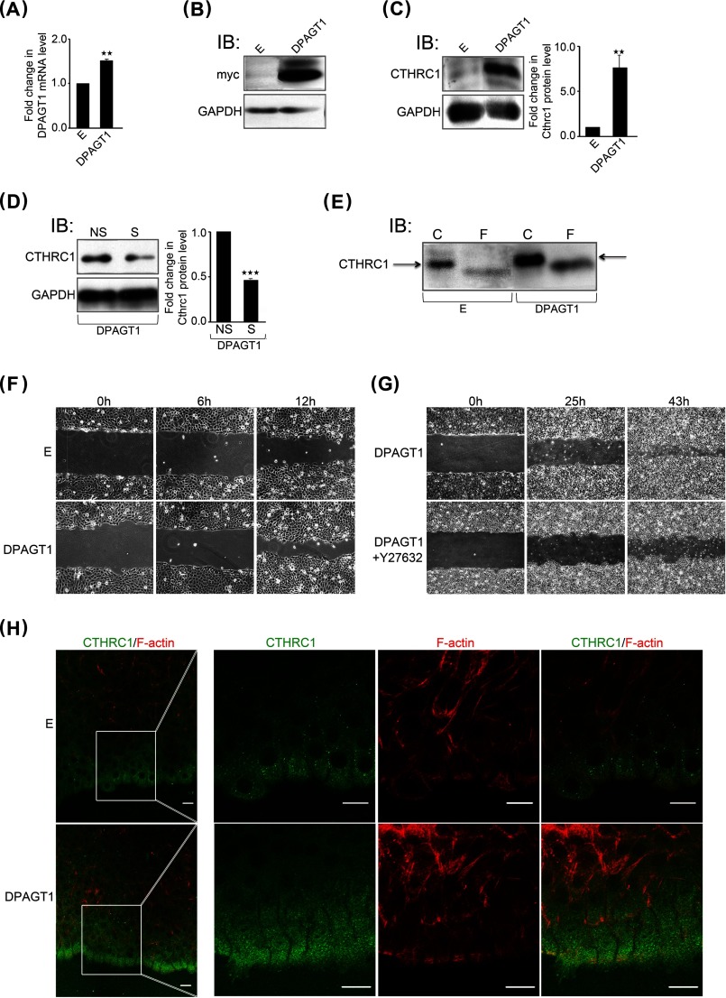
FIGURE 4.
Overexpression of DPAGT1 is associated with increased cell migration and augmented levels and localization of CTHRC1 to the wound front. A, quantitative PCR of DPAGT1 transcript levels in control empty vector (E)- and DPAGT1 cDNA-transfected CAL27 cells. **, p < 0.01. B, immunoblot (IB) of recombinant GPT isoforms (Myc tag) from empty vector and DPAGT1 transfectants. C, left, immunoblot of CTHRC1 from empty vector and DPAGT1 transfectants. Right, -fold change in CTHRC1 levels after normalization to GAPDH. **, p < 0.01. D, left, immunoblot of CTHRC1 from DPAGT1 transfectants treated with either NS or S (DPAGT1) siRNA. Right, -fold change in DPAGT1 levels after normalization to GAPDH. ***, p < 0.001. E, immunoblot of control (C) or PNGase F (F)-treated CTHRC1 from empty vector and DPAGT1 transfectants. F, scratch wound assay of empty vector- and DPAGT1-transfected CAL27 cells at 0, 6, and 12 h (×20 magnification). G, scratch wound assay of DPAGT1-transfected CAL27 cells in the absence or presence of a ROCK inhibitor, Y-27632 (30 μm), at 0, 25, and 43 h (×4 magnification). H, immunofluorescence localization of CTHRC1 (green) and F-actin (red) at the edge of a wound in empty vector- and DPAGT1-transfected cells at 18 h.

