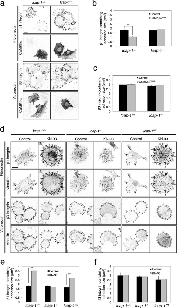
FIGURE 1.
CaMKIIα negatively regulates the formation of β1 integrin-dependent adhesion complexes in an ICAP-1α-dependent manner. a, Icap-1+/+ and Icap-1−/− osteoblasts expressing or not the constitutively activated CaMKIIαT286D and spread on FN (10 μg/ml) or on VN (5 μg/ml) for 2 h were immunostained to visualize β1 integrin (using 9EG7 antibody) and CaMKIIα. Scale bar, 10 μm. b and c, quantification of β1 (b) and β3 (c) integrin-containing FA size in Icap-1+/+ and Icap-1−/− osteoblasts expressing or not the constitutively activated CaMKIIαT286D. ***, p < 0.0001. d, Icap-1+/+, Icap-1−/−, and Icap-1WT osteoblasts spread on FN (10 μg/ml) or on VN (5 μg/ml) and treated or not with KN-93 (10 μm) for 2 h were immunostained to visualize vinculin and either activated β1 integrin (using 9EG7 antibody, for FN coating) or β3 integrin (for VN coating). Scale bar, 10 μm. e and f, quantification of β1 (e) and β3 (f) integrin-containing FA size in Icap-1+/+, Icap-1−/−, and Icap-1WT osteoblasts treated or not with KN-93 (10 μm) for 2 h. ***, p value < 0.0001. Error bars, S.D.

