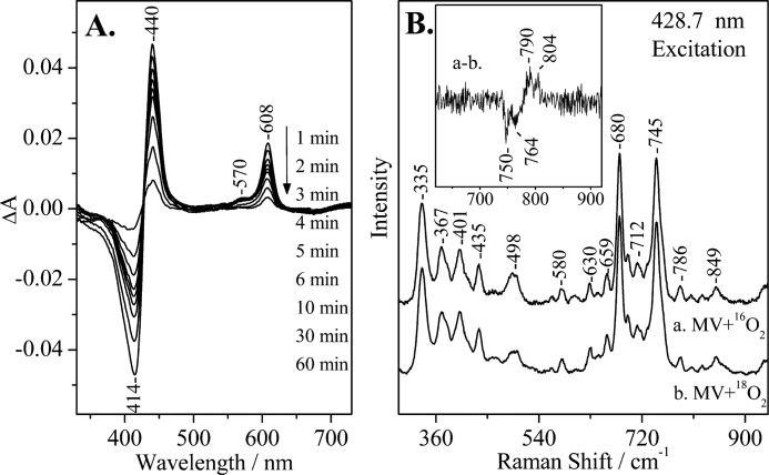
FIGURE 2.
A, optical absorption difference spectra of the reaction products by direct mixing of O2 with CO-MV (mixed valence) aa3 oxidase from P. denitrificans minus the oxidized form of the enzyme at the indicated times subsequent to mixing, pH 7.5. The enzyme concentration was 5 μm, and the path length of the cell was 0.5 cm. B, resonance Raman spectra of CO-MV at 0–5 min subsequent to mixing with 16O2 (spectrum a) and 18O2 (spectrum b). The inset shows the difference a-b spectrum. Several 0–5-min spectra were collected and added for the 16O2 and 18O2 experiments. The enzyme concentration was 50 μm, pH 7.5. The excitation wavelength was 428.7 nm, and the incident power was 1 milliwatt.

