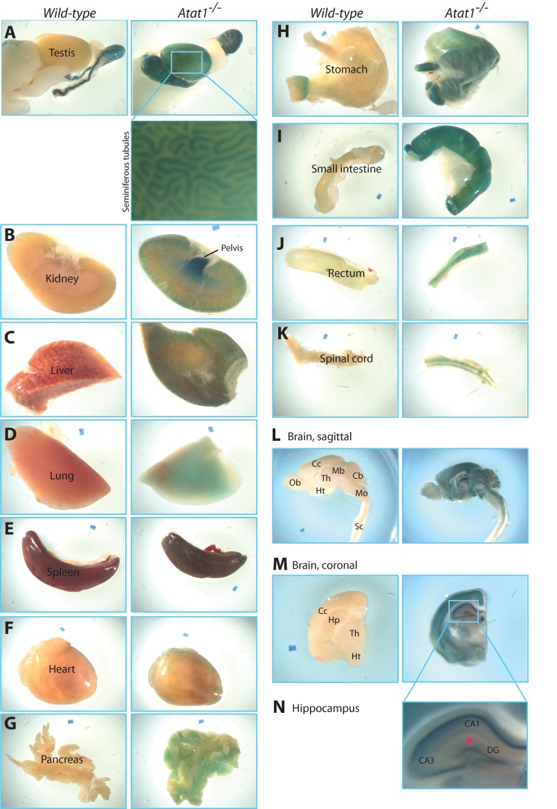
FIGURE 4.
Whole-mount β-galactosidase staining of adult mouse organs. A–N, strong expression was observed in the testis, renal pelvis, gastrointestinal tract, and brain of the Atat1−/− but not wild-type mouse. For the testis (A) and brain (M and N), two boxed regions are shown at higher magnifications to illustrate β-galactosidase activities detected in seminiferous tubules and the hippocampus of Atat1−/− mice. Although not clear from L, M, and N, high β-galactosidase activities were detected in the habenula as well as in islands of Calleja located within the olfactory tubercle. Different structures in the brain were labeled according to published atlases (71, 72). Abbreviations for regions marked in L, M, and N are as follows: CA1 and CA3, cornu ammonis areas 1 and 3 of the hippocampus, respectively; DG, dentate gyrus; Cc, cerebral cortex; Cb, cerebellum; Mb, midbrain; Mo, medulla oblongata; Hp, hippocampus; Ht, hypothalamus; Ob, olfactory bulb; Sc, spinal cord; Th, thalamus.

