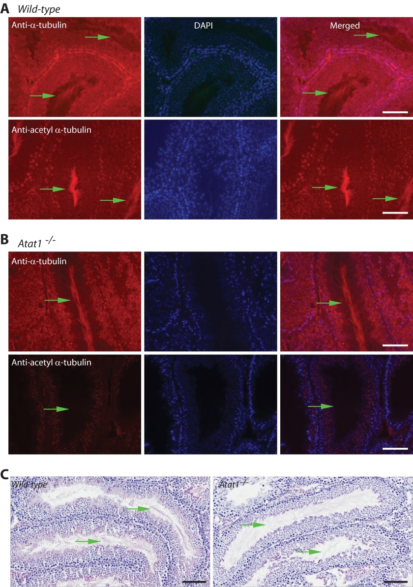
FIGURE 6.
Comparison of testis sections from wild-type and Atat1−/− mice. A and B, immunofluorescence microscopic analysis with antibodies recognizing regular (A) and acetylated (B) α-tubulin. Probing with the anti-α-tubulin antibody detected α-tubulin in almost every cell within seminiferous tubules of wild-type and Atat1−/− testis sections. In addition, strong signals were present at the luminal centers of the tubules (denoted with green arrows; A and B, top rows of images). The centers correspond to the areas where sperm flagella reside. Staining with the antibody against acetylated α-tubulin revealed the presence of α-tubulin acetylation at the central regions of seminiferous tubules in the wild-type testis section (A, bottom three images). Importantly, this signal was no longer observed in the testis section of the Atat1−/− mouse (B, bottom three images). Scale bar, 40 μm. C, H&E staining of testis sections. The sections were fixed in Bouin's solution and subjected to H&E staining. Green arrows denote sperm flagella. Scale bar, 100 μm.

