
FIGURE 8.
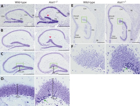
Histological analysis of wild-type and Atat1−/− hippocampi. A–C and E, representative hippocampal images taken from serial sections on four different sagittal planes in the medial-to-lateral direction. Nissl staining was performed as in Fig. 7, the two sections of which correspond to a sagittal position between those in A and B shown here. Red arrowheads in A and B mark the bulge in the lateral or suprapyramidal blade of the mutant dentate gyrus. Note that at the sagittal plane corresponding to that shown in Fig. 5D (middle and right), no bulge was found. D and F, high resolution images of the boxed areas in C and E, respectively. Green arrows denote darkly stained cell populations within the subgranular zone of the mutant dentate gyrus. D, the peak width of the granular cell layer is shown; the width was measured with the ruler tool of the Spectrum software used to analyze images digitized by the ScanScope. Abbreviations used are as follows: CA1 and CA3, cornu ammonis areas 1 and 3 of the hippocampus, respectively; DG, dentate gyrus; gcl, granular cell layer of the dentate gyrus; pyl, pyramidal layer of CA1 or CA3; SB, subiculum. Scale bar, 500 μm.