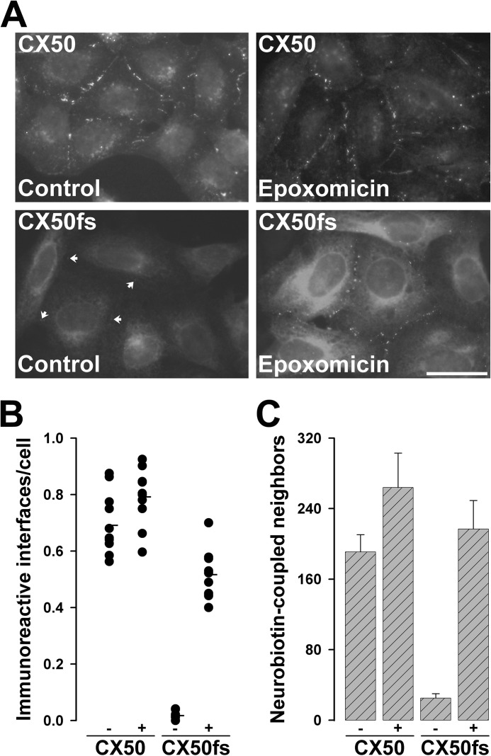
FIGURE 5.
Treatment of HeLa-CX50fs cells with epoxomicin increased gap junction plaques and rescued intercellular communication. A, photomicrographs show the distribution of CX50 and CX50fs in cells that were treated with DMSO (Control) or with 2 μm epoxomicin for 4 h. Arrows point to gap junction plaques between untreated HeLa-CX50fs cells. For either HeLa-CX50 or HeLa-CX50fs cells, the images of DMSO-treated and epoxomicin-treated cells were obtained with the same exposure time. Scale bar, 34 μm. B, the vertical point graph shows the quantification of the results presented in A. For each treatment, the total number of cell interfaces with immunopositive gap junction plaques and the total number of cells were counted in 10 different fields of view (containing confluent, but well spread cells). The results are presented as the number of immunoreactive interfaces per cell. The average for each treatment is indicated by the short horizontal line. C, the bar graph shows the quantification of intercellular transfer of Neurobiotin in HeLa-CX50 and HeLa-CX50fs cells after DMSO (−) or epoxomicin (+) treatment. The results are presented as mean + S.E. (error bars; p < 10−5 for untreated CX50fs versus CX50 or epoxomicin-treated CX50fs).

