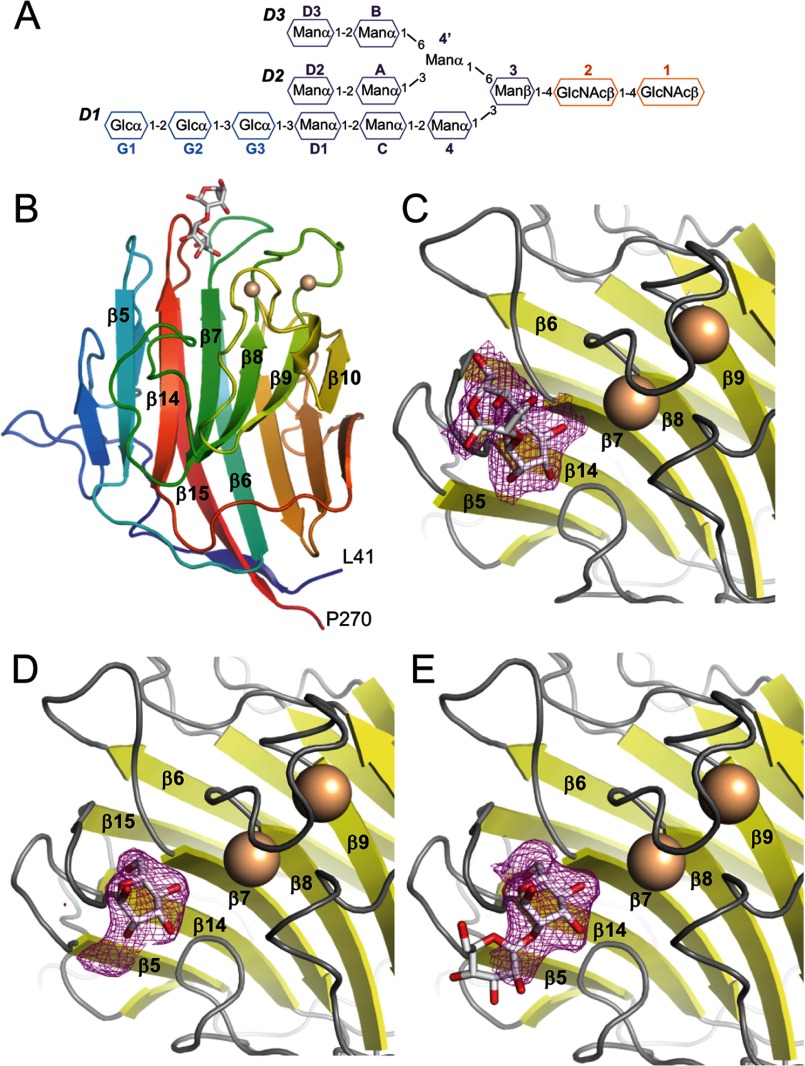
FIGURE 1.
Crystal structures of the LMAN1-CRD bound to Ca2+ and Man-α-1,2-Man. A, schematic representation of the glucosylated form of high mannose N-linked glycans, Glc3Man9(GlcNAc)2, added to secreted and membrane glycoproteins in the endoplasmic reticulum. B, overall structure of the LMAN1-CRD in the P1 crystal form; the P6 form of the protein is very similar. The two bound Ca2+ ions are shown as beige spheres, and Man-α-1,2-Man is shown in stick representation. Key secondary structure elements involved in glycan and calcium binding are labeled. C, Fo − Fc omit map density corresponding to the bound Man-α-1,2-Man in the P1 crystal, contoured at 2σ. D, Fo − Fc omit density corresponding to the bound Man-α-1,2-Man in the P6 crystal, contoured at 2σ. The density allows only one mannose group to be built. E, the “extra” density in the P6 crystal form may correspond to part of a mobile second mannose.

