FIGURE 3.
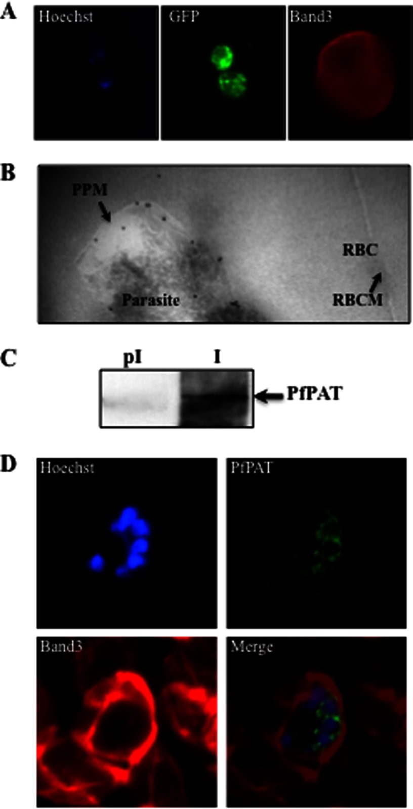
PfPAT localization in P. falciparum-infected erythrocytes. A, localization of PfPAT-GFP by immunofluorescence analysis using anti-GFP (green). The red blood cell membrane marker Band3 (red) was detected using an anti-Band3 monoclonal antibody. The parasite nucleus was visualized using the Hoescht 33258 dye (blue). B, transmission electron micrograph of ultrathin cryosections of the intraerythrocytic early trophozoite stage of P. falciparum PfPAT-GFP transgenic parasites using anti-GFP antibody (18-nm gold particles; indicated with arrows). PPM, parasite plasma membrane; RBC, red blood cell; RBCM, red blood cell membrane. C, detection of native PfPAT using affinity-purified PfPAT antibodies (I). Preimmune serum (pI) is used as a control. D, localization of PfPAT in P. falciparum 3D7 parasites at the schizont stage using anti-PfPAT antibodies (green). The red blood cell membrane marker Band3 (red) was detected using an anti-Band3 monoclonal antibody. The parasite nucleus was visualized using the Hoescht 33258 dye (blue).

