FIGURE 6.
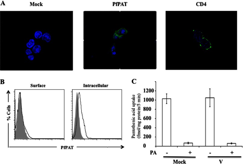
A, confocal microscopy of HEK-293T cells transfected with PfPAT-FLAG (middle) and stained with anti-FLAG FITC (green) or transfected with CD4-GFP as a control (right). Cell nucleus was visualized by staining with Hoescht 33258 dye (blue). B, flow cytometric analysis of HEK-293T cells mock transfected (gray) or transfected with PfPAT-FLAG (black) and surface stained or permeabilized for intracellular staining with anti-FLAG FITC. C, uptake of [3H]pantothenic acid (PA) by ARPE19 cells transiently transfected with pCMV-3Tag vector alone or with PfPAT cloned in the vector examined 48 h after transfection. Mock denotes the endogenous pantothenic acid transport by ARPE19 cells. The presence of carrier was confirmed by competing the [3H]pantothenic acid uptake by excess of unlabeled pantothenic acid. The data shown are means ± S.E. of five independent sets of experiments.

