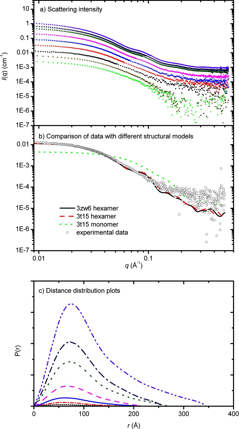
FIGURE 4.
X-ray scattering data of Rubisco activase. a, data were collected for tobacco Rubisco activase for a range (0.6–76 μm) of protein concentrations (top panel). b, data for 2.4 μm Rubisco activase overlaid with the scattering profiles, calculated using CRYSOL, for the open hexamer structure (generated from 3T15), closed hexamer structure (3ZW6) and monomeric structure (3T15). c, distance distribution functions, pr were determined using the indirect Fourier transformation package GNOM (bottom panel).

