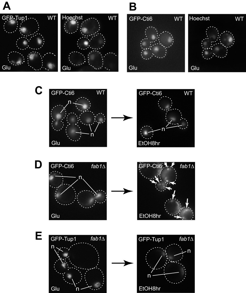
FIGURE 6.
Cti6 mislocalizes to the cytoplasm in fab1Δ cells in ethanol medium. A and B, GFP-Tup1 (A) and GFP-Cti6 (B) localizes primarily in the nucleus (stained with Hoechst 33342) in WT cells in glucose-rich medium. C, GFP-Cti6 localizes primarily in the nucleus in WT cells at 8 h after shifting from glucose to ethanol media. D, GFP-Cti6 accumulates in the cytoplasm in fab1Δ cells at 8 h after shifting from glucose to ethanol media. Some cytoplasmic Cti6 formed multiple speckles (marked by arrows). E, GFP-Tup1 localizes primarily in the nucleus in fab1Δ cells at 8 h after shifting from glucose to ethanol media. Single, bright GFP signals of Cti6 (C and D) or Tup1 (E) correspond to nuclear proteins (marked as n).

