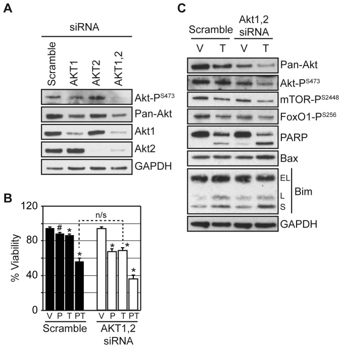
Figure 5. AKT depletion enhances tamoxifen cytotoxicity comparable to HDAC inhibition.
MCF7 cells were transfected with 1 µM scramble, AKT1, AKT2, or AKT1 and 2 directed siRNA for (A) 48 hours and western blotted or (B) 24 hours followed by treatment with vehicle (V), 0.1 µM PCI-24781 (P), 10 µM OH-tamoxifen (T), or the combination (PT) for an additional 72 hours and evaluated for cell viability. (C) MCF7 cells were transfected with 1 µM AKT1 and 2 directed siRNA for 24 hours, then divided and treated with vehicle (V) or 10 µM OH-tamoxifen (T) for 72 hours and western blotted. Cell viability treatments were conducted in triplicate with results expressed as the average with the error bars indicating the standard error of the mean. An (*) indicates a significant difference (P-value < 0.05) and a (#) an insignificant difference (P-value > 0.05) compared to vehicle treatment. n/s indicates an insignificant difference between treatments (P-value > 0.05).

