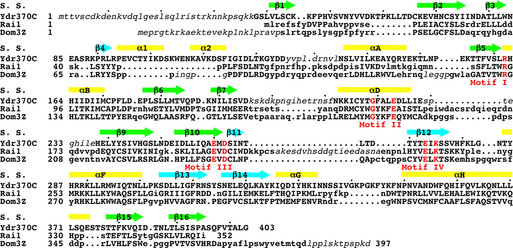
Figure 1.
Sequence conservation among Ydr370C/Dxo1, Rai1, and Dom3Z. Structure-based sequence alignment of K. lactis Ydr370C, S. pombe Rai1, and mouse Dom3Z. The secondary structure elements in the Ydr370C structure are shown (S. S.), and the four conserved sequence motifs are shown in red and labeled. Residues in Rai1 and Dom3Z that are located with 3 Å of the equivalent residue in Ydr370C are shown in uppercase. Residues that are disordered in the structures are shown in italic in lowercase.

