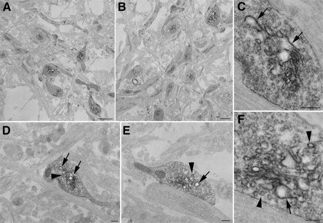
Figure 2.
FRET-based fluorescent photo-oxidation of ReAsH-labeled AS proteins expressed in neurons specifically labels membranous structures in the presynaptic terminals. A–F, Neurons were transfected with AS-EGFP-4Cys, cultured for 17 days, labeled with ReAsH, and FRET photo-oxidized to avoid nonspecific photo-oxidation of background staining. The EM staining reveals both large tubular membranous structures (arrows) and smaller vesicular structures (arrowheads). C and D are the same terminal, as are E and F. Scale bars, 2 μm (A), 1 μm (B), 200 nm (C–E), 100 nm (F).

