
Figure 8.
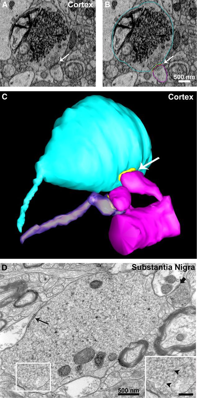
Atypical presynaptic terminals in neocortex and substantia nigra of AS transgenic mice. A–C, Using SBEM we observed, in the neocortex of AS transgenic mice, enlarged nerve terminals massively filled with an endomembrane network formed of tubulovesicular structures similar to those observed in hippocampus CA1. In C, three-dimensional volume segmentation shows the enlarged terminal (light blue) forming a synapse (postsynaptic density is in yellow, indicated by the white arrow) with a dendritic spine (magenta) compared to a normal terminal (purple) forming a synapse with an adjacent spine on the same dendrite. D, Similarly, TEM analysis of thin sections from substantia nigra of AS transgenic mice revealed endomembrane perturbations in presynaptic terminals characterized by extensive tubulovesicular structures (arrowheads in inset at higher magnification, scale bar, 250 nm). Slim black arrow points at a synapse formed by the enlarged terminal, while the shorter black arrow points at a synapse formed by a normal terminal.