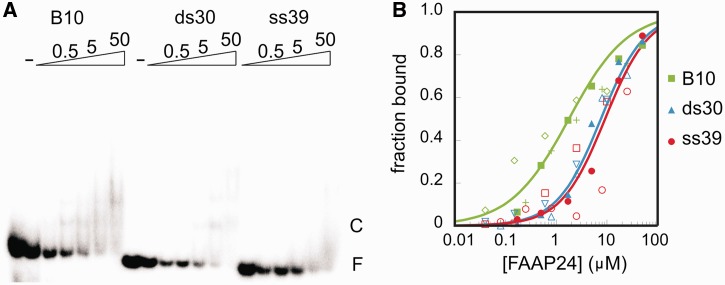
Figure 2.
Nucleic acid binding of the HhH domain of FAAP24. (A) Representative EMSA with a 39 nucleotides ssDNA (ss39), a 30 bp dsDNA (ds30) and a 30 bp bubble DNA (B10) consisting of two 10 bp dsDNA stems separated by 10 unpaired nucleotides. This binding assay is performed in the presence of 0 (−), 0.2, 0.5, 1.7, 5, 16.7, 50 μM FAAP24 HhH domain protein. The complex is marked as ‘C’, the free DNA as ‘F’. (B) Fraction bound DNA is plotted as a function of the FAAP24 HhH domain protein concentration. Experiments were performed in triplicate using B10 (green), ds30 (blue) ss39 (red); for each individual binding experiment, a different symbol is used, and the line represents the calculated binding curve based on the quantified apparent dissociation constant based on all data points, R2 of 0.9, 0.98, 0.92 for B10, ds30 and ss39 respectively.

