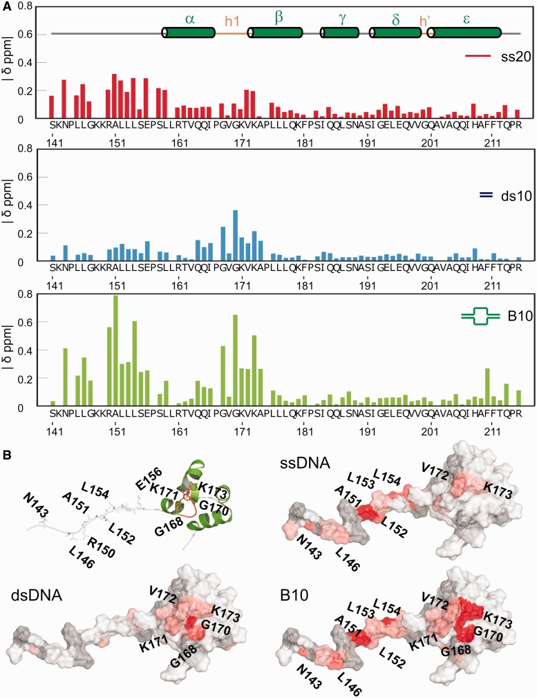
Figure 3.
The ssDNA and dsDNA interaction surfaces of the FAAP24 HhH domain. (A) NMR CSPs of 100 μM FAAP24 HhH domain protein with different DNA substrates. Compound CSP values were calculated as described before (16) and correspond to the addition of 150 μM ssDNA (ss20, red), 200 μM of dsDNA (ds10, blue) or 150 μM bubble DNA (B10, green). Secondary structure elements and hairpin regions are depicted in the top panel. (B) Surface representation of the FAAP24 HhH domain with CSPs on addition of the various DNA substrates plotted on the surface from white [composite CSP (ppm) <0.10; <0.10; <0.15 for ss20, ds10 and B10, respectively] to red (>0.3; >0.3: >0.4 for ss20, ds10 and B10, respectively). Gray indicates residues that could not be assigned or signals that disappeared in the titration.

