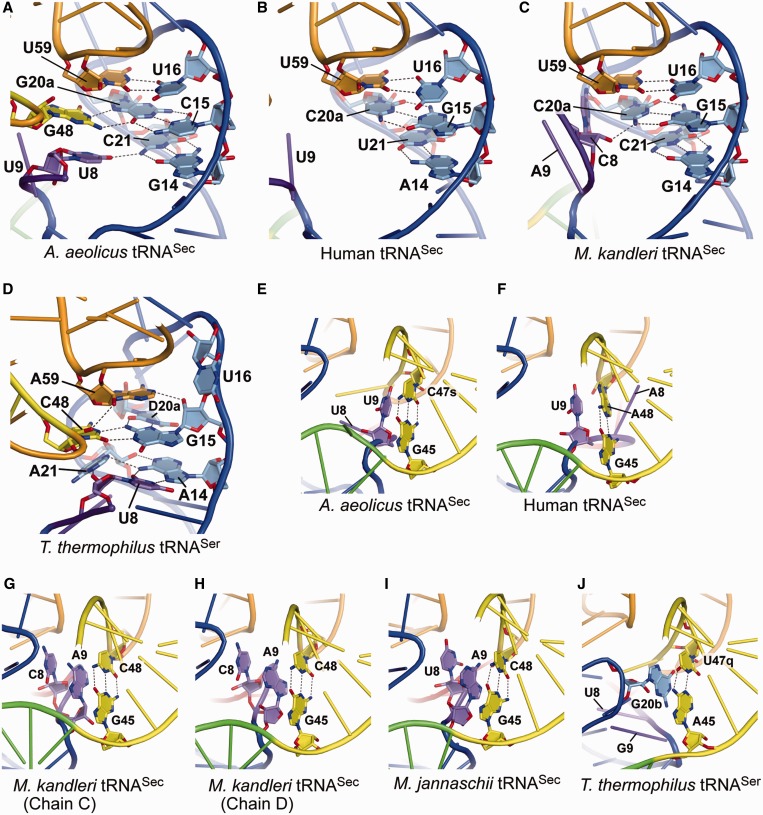
Figure 4.
Tertiary interactions. (A–D) Close-up views of the tertiary interactions with the AD linker and the D arm. The linker nucleotides U8 and G48 in A.aeolicus tRNASec interact with G14:C21 and C15:G20a in the D stem, respectively, to form unique base triples (A). In contrast, the D stem of human tRNASec lacks the tertiary interaction (B), while C8 in M. kandleri tRNASec interacts with the sixth D-stem base pair, G15:C21 (C). The N4 and O3′ atoms of C8 hydrogen bond with the O5′ and N4 atoms of C21, respectively. The nucleotides A14–A21 in T. thermophilus tRNASer are included in the D loop, and form the canonical tertiary pairs U8:A14 and G15:C48 (D). Furthermore, U8:A14 interacts with A21, and G15:C48 interacts with D20a and A59, thus building a tertiary core. (E–I) The tertiary stacking of the linker nucleotide at position 9 on the first extra-arm stem for the A. aeolicus (E), human (F), M. kandleri (G and H) and M. jannaschii (I) tRNASecs. Two distinct A9 conformations are observed in the two M. kandleri tRNASec molecules in the co-crystal with PSTK [PDB ID: 3ADD (11)]. The asymmetric unit contains two tRNASec molecules (chains C and D) and one PSTK dimer, and the A9s in chains C and D adopt different ribose puckering modes, C3′-endo and C2′-endo, respectively (G and H). The C2′-endo puckering is assumed by the U9s of the A. aeolicus (E) and human (F) tRNASecs. On the other hand, the asymmetric unit of the M. jannaschii tRNASec•PSTK co-crystal contains one tRNASec molecule and one-half of a PSTK dimer [PDB ID: 3AM1 (14)], and the A9 ribose puckering is C2′-endo (I). C8 and U8 of the M. kandleri and M. jannaschii tRNASecs, respectively, are further stacked on A9. (J) The tertiary stacking in T. thermophilus tRNASer. The D-loop nucleotide G20b is stacked on the first base pair of the extra-arm stem.

