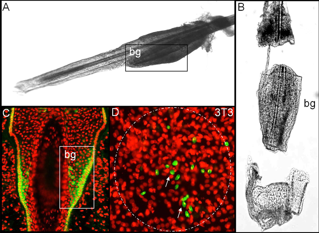
Figure 1.
Stem cell isolation and expansion (A, B) Murine vibrissae hair follicles were isolated under a dissecting microscope, in order to access the bulge region harbouring the epithelial stem cells (SC), which express keratin 15 (Krt15) (C), the mesenchymal capsule was completely removed. (D) Following the enzymatic digestion of the epithelial core´s central portion, the cells were seeded on a 3T3 feeder cell layer to purify and enrich the SC by means of clonal expansion. The presence of the putative SC marker Krt15 (green) was confined to several small cell clusters (arrow) within each holoclone. bg: bulge; nuclear staining: propidium iodide (red). A,B,C: reprinted with permission [19].

